Scientist Found Changes Identified In The Brains Of Migraine Patient

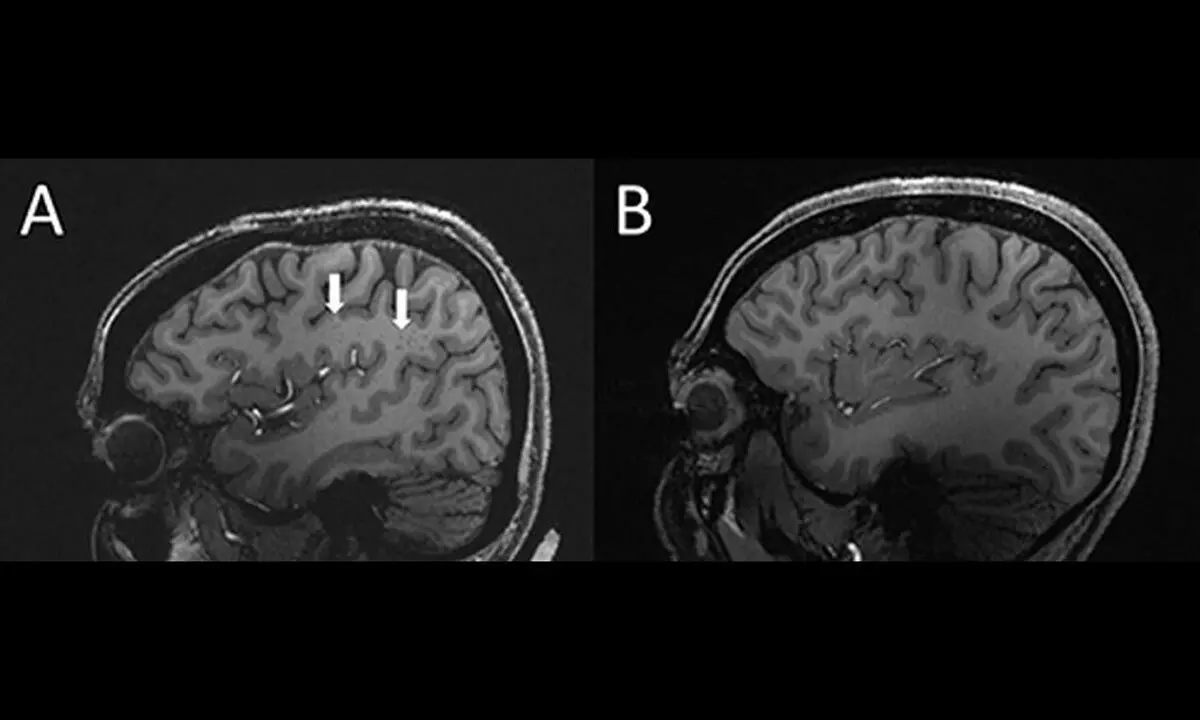
The arrows show enlarged perivascular spaces in the brain of a chronic migraine patient (left) compared to a healthy control (right). (Radiological Society of North America)
- The vexing and enduring enigma of the migraine may have just been solved due to the new clue discovered by scientists.
- Researchers discovered that patients who suffer from episodic and chronic migraine had significantly enlarged perivascular gaps, the fluid-filled regions around the brain's blood arteries.
The vexing and enduring enigma of the migraine may have just been solved due to the new clue discovered by scientists. Researchers discovered that patients who suffer from episodic and chronic migraine had significantly enlarged perivascular gaps, the fluid-filled regions around the brain's blood arteries.
The discovery might represent a new study direction that has not yet been explored, even though the connection to or role in migraine has not yet been determined. The discovery was announced at the Radiological Society of North America's 108th Scientific Assembly and Annual Meeting.
Ten percent of the world's population is thought to be impacted by the ailment. Therefore, identifying a cause and developing better management techniques would improve millions of people's lives.
The perivascular areas in the centrum semiovale, the primary area of brain white matter directly beneath the cerebral cortex, piqued the interest of Xu and his coworkers. Although the purpose of these areas is not fully known, they do contribute to fluid outflow and their expansion may be a sign of more serious issues.
He and his colleagues gathered 20 migraine sufferers between the ages of 25 and 60: 10 of whom had chronic migraine without aura and 10 of whom had episodic migraine. A control group of 5 healthy patients who don't get migraines was also added.
Patients with cognitive impairment, claustrophobia, brain tumours, or previous brain surgery were disqualified by the researchers. Then, they used an ultra-high-field MRI using a 7-tesla magnet to perform MRI images. The majority of hospital scanners only use 3 tesla magnets. The scans showed that migraine patients' perivascular spaces in the centrum semiovale were substantially larger than those of the control group.
Additionally, white matter hyperintensities, a particular kind of lesion, were discovered to be distributed differently in migraine sufferers. These are rather typical and are brought on by little areas of dead or partially dead tissue that are starved by reduced blood flow.
The frequency of these lesions was the same in both migraine patients and control patients, but the severity of deep lesions was greater in migraine patients. This suggests, in the opinion of the researchers, that the expansion of perivascular spaces may eventually result in the development of additional white matter lesions.
The results indicate that migraine is associated with a problem with the brain's plumbing, specifically the glymphatic system, which is in charge of clearing waste from the brain and nervous system, even though the nature of the connection between enlarged perivascular spaces and migraine is unclear. It transports through perivascular channels. However, even though further research is needed to fully understand this association, its discovery is encouraging.
Next Story


